


The main costochondritis symptoms are sharp, aching or pressure-like pain at the front of the chest, beside the breastbone, often over several rib joints, which pressing on the spot reproduces. It gets worse with deep breaths, coughing, lying down and moving your upper body. Reproducible pain is reassuring, but it does not rule out the heart.
If you are reading this, you have probably pressed on your chest and found the pain under your finger. That is reassuring. It is not proof. Here is how to tell what you have, how long it really lasts, and what eases it.
What are the symptoms of costochondritis?
Your ribs do not attach straight onto the breastbone. Between the bone of each rib and the sternum sits a length of cartilage, the costal cartilage, which gives the ribcage the flexibility to lift with every breath. Costochondritis is inflammation where those cartilages meet the ribs or the breastbone.
The NHS describes sharp pain at the front or side of the chest that gets worse when you move your upper body, lie down, breathe deeply, or press on the middle of your chest or your ribs. Many people also feel it when they cough, sneeze, lift their arms or carry shopping. The pain can be stabbing, a dull ache or a feeling of pressure, and it often sits over several junctions rather than one point.
Where it hurts follows a pattern. In a German study of 1,212 people aged 35 and over who saw their GP with chest pain, the pain came from the chest wall in 46.6% of cases, making it the commonest cause in general practice. In those people the pain sat behind or beside the breastbone in 52.0% and on the left side in 69.2%, which explains why so many fear their heart.

Costochondritis is also more common in women. In a New York emergency department, 69% of the patients diagnosed with it were women, against 31% of those whose chest pain had another cause. The symptoms themselves are the same in women and men.
Why do I suddenly have costochondritis?
Often nobody finds a single cause. The NHS links costochondritis to a chest injury, repetitive upper-body movements such as lifting, a lot of coughing, and infection. A chest cold that leaves you coughing for weeks, a weekend of moving furniture or a new gym programme heavy on push-ups are typical starting points.
It is a real inflammation, not imagined pain, and imaging has shown it in the swollen form. In Siena, a team scanned 12 people with Tietze syndrome by MRI: the cartilage was thickened in all 12, it took up contrast in 10, and the bone next to it was swollen with fluid in 5. A blood test can show slightly raised inflammation markers too, although in the New York study the average sedimentation rate was no different from that of other chest pain patients.
Costochondritis or Tietze syndrome: what is the difference?
Both affect the same cartilages, and plenty of clinicians use one word for the other. One sign separates them, and you can check it yourself: swelling.
Costochondritis is pain at the rib and breastbone junctions with no visible swelling. It is very common: in the New York emergency department it accounted for 36 of 122 patients with chest pain, or 30%. Tietze syndrome, described in 1921 by the German surgeon Alexander Tietze, is rare, and it swells. A firm, tender lump forms over one junction, usually the second or third, on one side only, and the skin over it can feel slightly warm.
| Feature | Tietze syndrome | Costochondritis | Intercostal neuralgia |
|---|---|---|---|
| Swelling | Yes, firm, over the junction | No | No |
| Painful area | Usually a single point | Often several points | A band along one rib |
| Usual site | 2nd or 3rd rib, at the edge of the breastbone | Edge of the breastbone, often from the 2nd to the 5th rib | From the back round to the front, on one side |
| Type of pain | Dull, sharp when pressed | Sharp when pressed and when twisting | Burning, electric shocks |
| What sets it off | Pressure, coughing, turning the trunk | Pressure, deep breaths, effort with the arms | Coughing, sneezing, clothes rubbing |
| Tissue involved | Inflamed cartilage | Cartilage and junction, without swelling | The nerve running under the rib |
Intercostal neuralgia involves a different tissue: the nerve tucked under each rib is irritated, not the cartilage. Its causes and treatments differ, and they are covered in our guide to intercostal neuralgia.

How is costochondritis diagnosed? The press test
Diagnosis starts with the examination. Your GP looks for the exact pain under a fingertip, and you can repeat the move at home, sitting in front of a mirror with your top off.
The press test, in four steps
Find the landmark
Follow the breastbone down from the notch at the base of your neck. A few centimetres lower, a small horizontal ridge marks where the second rib attaches.
Press joint by joint
With two fingers, press firmly along the edge of the breastbone, rib by rib, from top to bottom, on the left and then on the right.
Compare with your pain
The pressure should reproduce exactly the pain that worries you, in the same place, not just a vague tenderness.
Look for a lump
Compare both sides by eye and with your fingertips. A firm bump over a single junction suggests Tietze syndrome; pain without a bump over several points, costochondritis.
An ultrasound scan, quick and free of radiation, can then show a thickened cartilage, and an MRI also picks up inflammation in the neighbouring bone. A chest X-ray is usually normal, because cartilage does not show on it: its job is to rule out a lung problem.

A review in American Family Physician sums up the rule doctors follow: in children, teenagers and young adults, pain reproduced by pressing on the cartilages is usually all that is needed. Anyone over 35, with heart disease or its risk factors, or with breathing or heart symptoms should have an ECG and sometimes a chest X-ray.
Chest pain: when should you not wait?
This is the most important part of the article. Pain you can find under your finger is reassuring, often rightly so. It is not enough on its own.
In the New York emergency study, 6% of patients with costochondritis were having a heart attack at the same time. The American Family Physician review puts coronary disease at 3 to 6% of adults with chest pain and a tender chest wall. A review in JAMA points the same way: pain that is stabbing, positional or reproducible by pressure makes a heart problem less likely, with likelihood ratios of 0.2 to 0.3, but no part of the history is strong enough on its own to skip testing.
Heart attacks among 122 emergency patients with chest pain
The NHS asks you to call 999 for chest pain that started suddenly, spreads to your arm, neck or jaw, or makes your chest feel tight, which could be a heart attack. Call 999 too for chest pain with difficulty breathing, a racing heart or coughing up blood, which could be a clot in the lung, and for sudden chest or back pain that feels like tearing inside. Do not drive yourself to A&E.
If you think you have costochondritis and none of those signs apply, the NHS still advises getting help from 111, because unexplained chest pain should always be checked. Book your GP promptly if you are over 35, have heart risk factors or a heart history, even when the pain sits under your finger. An ECG takes a few minutes. A fever with a cough and pain on breathing in points instead to the lungs or the lining around them.
How long does costochondritis pain last?
Often longer than people hope. The NHS says it can last from a few weeks to several months, and the few follow-up studies agree.
In the German GP study, 55.4% of people with chest wall pain still had chest pain six months later. In the New York study, 11 of the 21 patients seen again after a year were still in pain, although only a third of them still had clear costochondritis. The authors concluded that most cases settle on their own within a year.
Tietze syndrome also heals by itself, without after-effects, but it is counted in weeks and sometimes months. What drags both out is mostly going on loading the junctions: lifting, push-ups or bench press, an untreated cough, nights spent lying on your front.
How can costochondritis be treated?
No controlled trial has compared treatments for costochondritis or Tietze syndrome. Care rests on clinical practice and small case series, which is a good reason to distrust promises of a quick cure, including the many “how I cured my costochondritis” stories.
Rest the junction, not your whole body
For a few weeks, cut back on what hurts: heavy lifting, pushing, pulling, push-ups. Get a lingering cough treated, because every fit of coughing pulls on the cartilage. Sleep on your back or on the side that does not hurt, and keep walking and moving normally.
Cold calms a warm, swollen spot; heat relaxes the tense muscles around it. If you are torn between the two, our guide to ice or heat sets out how to choose, and the same logic applies here.
Painkillers
The NHS mentions anti-inflammatory painkillers such as ibuprofen; paracetamol, or an anti-inflammatory gel rubbed on the area, are alternatives. Take them for short periods, and ask a pharmacist first if you have a sensitive stomach, kidney disease or take heart medicines.
An injection for pain that will not settle
For very bad pain that does not improve, the NHS says you may be offered a steroid injection or a local anaesthetic into the junction. In an Egyptian series of 9 patients with Tietze syndrome, the cartilage had shrunk on ultrasound one week after the injection, and the procedure proved safe. Surgery to remove the diseased cartilage remains exceptional, kept for stubborn cases.
Supporting an inflammatory background
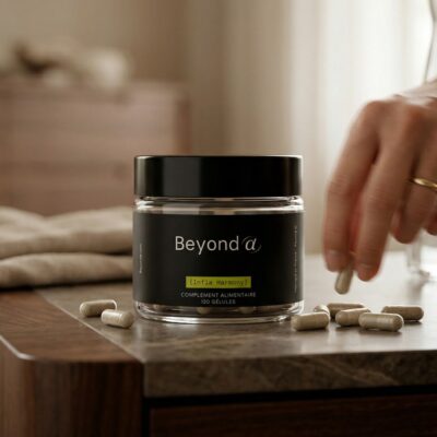
When the pain lasts for months, some people choose to add INFLA HARMONY, a formula for long-lasting pain combining curcumin, boswellia, ginger, quercetin and alpha-lipoic acid. It replaces neither medical advice nor rest for the junction. It is not recommended with anticoagulants, which many people with heart conditions take, with diabetes, a bile duct disorder, pregnancy or breastfeeding, or with cancer medicines or immunosuppressants.
When is it more than costochondritis?
True Tietze syndrome affects one junction and eventually heals. If the swelling lasts, keeps returning or involves several joints at the top of the chest, the diagnosis deserves a second look.
A Japanese review published at the end of 2024 points out that several inflammatory rheumatic diseases swell exactly the same joints: spondyloarthritis, rheumatoid arthritis, SAPHO syndrome, pustulotic arthro-osteitis and gout. Go back to your GP if you notice any of these:
- back pain that wakes you in the second half of the night and eases with movement, typical of inflammatory back pain;
- psoriasis, or pustules on the palms and soles;
- pain in a heel, a buttock or other joints;
- a fever, clear redness over the junction, or a hard lump that keeps growing.
A red, hot junction with a fever, especially after chest surgery, raises the fear of infection. A growing lump needs imaging: tumours of the rib cartilage are rare, but they exist.
Frequently asked questions
Can stress cause costochondritis?
Stress does not inflame the rib cartilage directly. It can tighten the chest and shoulder muscles, make you breathe shallowly and lower your pain threshold, so an existing costochondritis feels worse. Anxiety can also cause chest pain of its own. If you are unsure what is behind your chest pain, get it checked rather than putting it down to stress.
What should you not do with costochondritis?
For a few weeks, avoid what loads the rib joints: heavy lifting, push-ups, bench press, carrying heavy bags on one side and sleeping on your front. Get a lingering cough treated. Do not keep pressing the sore spot to check on it, and never assume that a new or different chest pain is just costochondritis.
Is costochondritis more common in women?
It seems to be. In an emergency department study of 122 people with chest pain, 69% of those with costochondritis were women, against 31% of the others. The symptoms are the same in women and men, and so are the warning signs that call for 999: pain spreading to the arm, neck or jaw, tightness or breathlessness.
Can costochondritis come back?
Yes. It usually settles over weeks to months, but flare-ups are common when the junctions are loaded again, after a cough, heavy lifting or a new exercise programme. In one study, 11 of 21 people still had chest pain a year later. A swelling that keeps returning, or several joints involved, deserves a check for inflammatory arthritis.
Sources
Disla E., Rhim H.R., Reddy A. et al. (1994). Costochondritis: a prospective analysis in an emergency department setting. Archives of Internal Medicine, 154(21), 2466-2469. DOI: 10.1001/archinte.154.21.2466
Bösner S., Becker A., Hani M.A. et al. (2010). Chest wall syndrome in primary care patients with chest pain: presentation, associated features and diagnosis. Family Practice, 27(4), 363-369. DOI: 10.1093/fampra/cmq024
Swap C.J., Nagurney J.T. (2005). Value and limitations of chest pain history in the evaluation of patients with suspected acute coronary syndromes. JAMA, 294(20), 2623-2629. DOI: 10.1001/jama.294.20.2623
Proulx A.M., Zryd T.W. (2009). Costochondritis: diagnosis and treatment. American Family Physician, 80(6), 617-620. PubMed: 19817327
Volterrani L., Mazzei M.A., Giordano N. et al. (2008). Magnetic resonance imaging in Tietze’s syndrome. Clinical and Experimental Rheumatology, 26(5), 848-853. PubMed: 19032818
Kamel M., Kotob H. (1997). Ultrasonographic assessment of local steroid injection in Tietze’s syndrome. British Journal of Rheumatology, 36(5), 547-550. DOI: 10.1093/rheumatology/36.5.547
Rokicki W., Rokicki M., Rydel M. (2018). What do we know about Tietze’s syndrome? Polish Journal of Cardio-Thoracic Surgery, 15(3), 180-182. DOI: 10.5114/kitp.2018.78443
Matsuki Y., Nakamura T. (2024). Spondyloarthritis and Tietze’s syndrome: a re-evaluation. Modern Rheumatology, 35(1), 1-6. DOI: 10.1093/mr/roae086


